Yes. Cataract surgery in Korea is often possible for people with macular degeneration, diabetic retinopathy or an epiretinal membrane, but a full retina exam comes first. The exam assesses how active the retinal disease is, the expected visual benefit and any need for additional treatment, and it helps guide three decisions: surgical timing, lens selection and the follow-up plan.
Medically reviewed by Dr. Park JinHeung, Director, St. Mary’s Jin Eye Center (Ophthalmologist, PhD; Adjunct Professor, Seoul St. Mary’s Hospital) · Last updated 2026-10-09
Cataract surgery replaces the cloudy natural lens with a clear artificial lens (intraocular lens). Light is then focused onto the retina, and its center, the macula, provides the detailed central vision needed for reading and recognizing faces. When the retina is not healthy, the operation itself is usually the same, but the plan around it changes. This guide explains what changes, based on published studies, and what overseas patients should prepare before traveling to Seoul.
Can I have cataract surgery if I have a retinal condition?
Usually, yes. The type and activity of the retinal condition decide the timing, and the final vision depends on the macula, retina and optic nerve, not only on the new lens.
A Cochrane review (2017) found two randomized trials (114 people) on cataract surgery in macular degeneration. In one of them, people operated on immediately had better corrected vision at six months than those whose surgery was delayed, but the reviewers concluded that the long-term benefit and harm remain uncertain. The American Academy of Ophthalmology (AAO) guideline also lists better management of coexisting retinal disease as one reason for cataract surgery.
Expectations matter. If the macula or optic nerve is already damaged, removing the cataract may not restore normal vision. That is why the retina is checked first and the expected result is discussed before surgery.
Why is a retina exam done before cataract surgery?
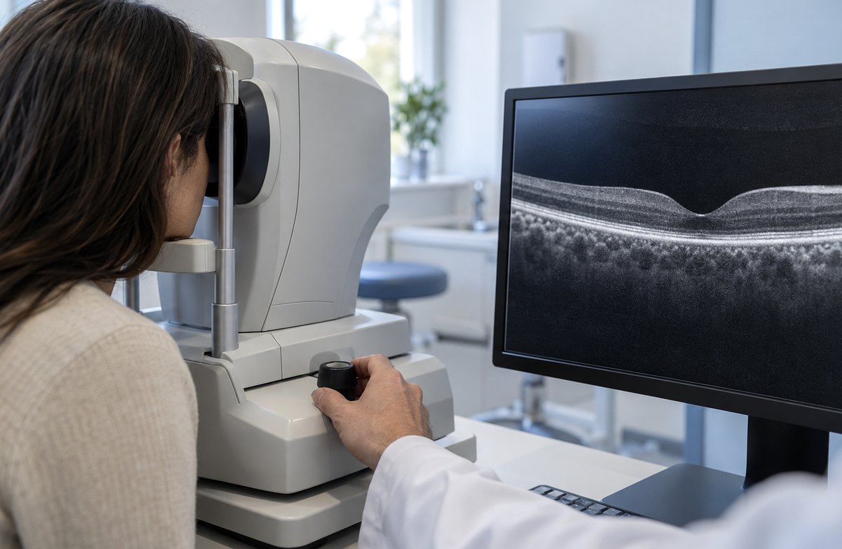
To find macular changes that a routine exam can miss. A dense cataract can hide them, and OCT can show fine structural changes even when the macula looks normal.
The AAO guideline includes a dilated exam of the lens, vitreous, macula, peripheral retina and optic nerve before surgery, and notes that OCT can help assess the central macula and coexisting retinal disease.
How often does OCT find something? In a Brazilian study presented at ASCRS 2018, 952 of 1,060 eyes scheduled for surgery looked normal on the routine exam; OCT found abnormalities in 4.9% of them, two-thirds of which were epiretinal membranes. In a 2026 Japanese study of 316 eyes, OCT detected previously unsuspected macular lesions in 30 eyes (9.5%), more often in older patients; three eyes (1.0%) had clinically significant findings requiring additional treatment or a change of the surgical plan. The rate depends on the age and selection of the patients.
| Test | What it shows | Why it matters for cataract surgery |
|---|---|---|
| Dilated fundus exam | Macula, peripheral retina and optic nerve | Retinal tears, bleeding, stage of diabetic retinopathy |
| Macular OCT | Cross-section of the macula, layer by layer | Epiretinal membrane, macular edema, macular degeneration; baseline for comparison after surgery |
| Fundus photography | A photo record of the retina | Tracking diabetic retinopathy and macular degeneration over time |
If the cataract is very dense, OCT image quality may be limited. If the back of the eye cannot be seen, an eye ultrasound (B-scan) helps detect major problems such as retinal detachment (it does not replace OCT for the fine structure of the macula), and macular OCT may be repeated after surgery.
What changes if I have macular degeneration?
Surgery is usually possible. Large studies have not shown a meaningful link between cataract surgery and progression to late macular degeneration. With wet macular degeneration, surgery is planned when the disease is controlled, in coordination with your injection treatment.
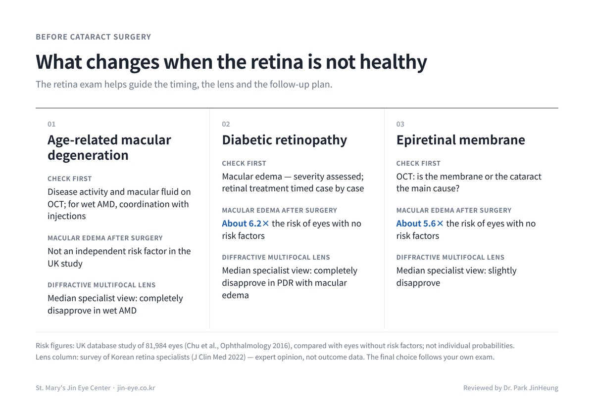
The US AREDS2 study (Ophthalmology 2022) analyzed 1,767 eyes that had cataract surgery during an average follow-up of about nine years and found no statistically significant increase in the risk of late macular degeneration. AREDS2 enrolled people already at risk of progression and analyzed eyes without late disease at the time of surgery, so the result cannot be applied directly to every patient with advanced disease. In the UK study of 81,984 eyes, macular degeneration was not an independent risk factor for macular edema after surgery. With wet (neovascular) macular degeneration, disease activity and macular fluid are assessed on OCT before surgery, and the operation is generally planned when the disease is adequately controlled, in coordination with ongoing anti-VEGF treatment. Regular retina checks continue afterwards.
If you receive anti-VEGF injections, send your treatment dates and recent OCT results so the surgeon can coordinate your care with your retina specialist.
What changes if I have diabetic retinopathy or an epiretinal membrane?
The risk of macular edema after surgery is higher, so the macula is checked first. If macular edema is present, its severity and activity are assessed, and retinal treatment may be recommended before, around or after cataract surgery, depending on the case.
In the UK study (Chu et al., Ophthalmology 2016), the rate of macular edema after surgery was 1.17% in eyes without complications, diabetes or other risk factors. Compared with those eyes, the risk was about 1.8 times higher with diabetes but no retinopathy, about 6.2 times higher with diabetic retinopathy (rising with severity) and about 5.6 times higher with an epiretinal membrane. Eyes receiving prophylactic nonsteroidal anti-inflammatory drugs (NSAIDs) were excluded from the study. These are relative risks, not personal probabilities.
| Condition | Relative risk of macular edema after surgery | Typical plan |
|---|---|---|
| No risk factors | Reference (rate 1.17%) | Routine follow-up |
| Diabetes, no retinopathy | About 1.8× | Dilated exam to confirm no retinopathy |
| Diabetic retinopathy | About 6.2× (higher if more severe) | Check for macular edema; treat the retina first if needed; anti-inflammatory drops may be considered |
| Epiretinal membrane | About 5.6× | OCT to judge severity and whether retinal treatment is also needed |
| Previous retinal vein occlusion | About 4.5× | Check the current macula and watch for edema after surgery |
| Uveitis | About 2.9× | Operate when the inflammation is quiet |
Source: Chu et al., Ophthalmology 2016 (UK, 81,984 eyes, retrospective; eyes receiving prophylactic NSAIDs excluded). Relative risks compare with eyes without risk factors.
With an epiretinal membrane, the key question is which problem limits your vision. If the membrane has little effect and the cataract is the main cause, cataract surgery can be done first and the macula watched. If the membrane is distorting the macula, cataract surgery alone may not give the expected result, and the need for retinal treatment and its order are discussed.
Can I have a multifocal lens with a retinal condition?
If the condition involves the macula, a diffractive multifocal lens is often not recommended, and a monofocal or other lens is discussed instead. Multifocal designs split light between focal points, which can be a disadvantage when the macula is weak.
In a survey of 111 Korean retina specialists (J Clin Med 2022), the median answer was “completely disapprove” of a diffractive multifocal lens for wet macular degeneration and for proliferative diabetic retinopathy with macular edema, and “slightly disapprove” for an epiretinal membrane (52.3% against). For lattice degeneration or a laser-treated retinal tear, more than 80% had no reservations. This is expert opinion, not a comparison of surgical results, and the final choice follows your own examination.
What should overseas patients plan for cataract surgery in Korea?
Allow time for the retina exam, the surgery and at least the day-3 check-up, ideally about one week in Seoul, send your retina records before you travel, and arrange local follow-up before you return.
| Step | At St. Mary’s Jin Eye Center |
|---|---|
| Before you travel | Email recent retina reports, OCT scans, fundus photos, injection/laser history and HbA1c (if diabetic) to jin.eye.center@gmail.com for a preliminary review |
| Exam day | Eye examination about 1 hour, consultation and consent about 1 hour; same-day surgery may be possible in selected cases; with active retinal disease, macular edema or findings that need additional treatment, separate visits or a revised schedule may be needed |
| After surgery | Rest and observation at the clinic for 30 minutes to 1 hour, then back to your hotel once the staff confirm you are ready |
| Stay | Some patients may be cleared to travel after the day-3 check-up if recovery is uncomplicated and the surgeon confirms it; about one week is recommended. Active retinal disease or additional retinal treatment may require a longer stay |
| Cost | Monofocal cataract surgery from KRW 1,500,000 per eye. The final quote is confirmed after the detailed examination and depends on the lens chosen |
Contact lenses: as a usual guide, stop soft lenses 5 days, hard (RGP) lenses 2 weeks and ortho-K lenses 4 weeks before the exam. Long-term rigid or ortho-K wearers may need longer, and final lens measurements are taken only once corneal readings are stable, so confirm your schedule with the clinic. Prices are confirmed after the detailed examination.
Macular edema after cataract surgery often develops several weeks after the operation, commonly around 4 to 6 weeks, though it can appear earlier or later; with diabetic retinal disease, existing macular edema can also worsen. This is often after you have returned home. Before leaving Korea, confirm a follow-up appointment with a local ophthalmologist or retina specialist, and know whom to contact if your vision changes.
When should I seek care urgently after surgery?
A curtain or shadow over your vision, or a sudden drop in vision, needs emergency eye care immediately. A sudden increase in floaters or flashes of light, new distortion or a central blind spot needs an eye exam the same day.
| Symptom after surgery | Possible cause | When |
|---|---|---|
| Vision becomes blurry again after first improving | Macular edema, inflammation, raised eye pressure or other complications | Same day; immediately (emergency) if sudden or severe |
| New or worse distortion, or a blank spot in the center | Macular edema, wet macular degeneration | Same day; immediately (emergency) if central vision loss is sudden or severe |
| Sudden increase in floaters, flashes of light | Retinal tear or detachment | Same day, without delay |
| A curtain or shadow over part of the vision | Retinal detachment | Immediately (emergency) |
| Severe pain, marked redness or sudden loss of vision (any one of these) | Infection (endophthalmitis) and others | Immediately (emergency) |
If you are already back home, go to an emergency eye service near you. Instructions from the surgeon who operated on you come first.
Frequently asked questions
Can I have cataract surgery if I have macular degeneration?
Usually, yes. In a large US study (AREDS2), cataract surgery was not associated with a statistically significant increase in the risk of progressing to late macular degeneration in the people studied; this cannot be applied directly to every patient with advanced disease. How much your vision improves depends on the condition of your macula, and if you have wet macular degeneration, surgery is planned when the disease is controlled, in coordination with your injection treatment.
What is the main risk of cataract surgery with diabetic retinopathy?
Swelling of the macula (macular edema) after surgery. In a UK study of 81,984 eyes, eyes with diabetic retinopathy had about 6.2 times the risk of eyes without risk factors. That is why the retina is checked before surgery. If diabetic macular edema is present, its severity and activity are assessed, and retinal treatment may be recommended before, around or after cataract surgery, depending on the case.
Why do I need an OCT scan before cataract surgery?
OCT shows fine structural changes in the macula that a routine eye exam can miss, and a dense cataract can hide them further. In a Brazilian study, OCT found abnormalities in 4.9% of eyes that looked normal on the routine exam; in a separate Japanese study, previously unsuspected macular lesions were found in 9.5% of 316 eyes examined before surgery (the two studies used different criteria and denominators). Depending on how significant they are, such findings may affect the timing, the lens choice or the follow-up plan.
Can I choose a multifocal lens if I have a retinal condition?
If the condition involves the macula, a diffractive multifocal lens is often not recommended, because it splits light between several focal points. In a survey of Korean retina specialists, the median answer was “completely disapprove” for wet macular degeneration and for proliferative diabetic retinopathy with macular edema. A monofocal or other lens is then usually discussed.
How long should I stay in Korea after cataract surgery?
At St. Mary’s Jin Eye Center, some patients may be cleared to fly home after the day-3 check-up, provided recovery is uncomplicated and the surgeon approves travel; a stay of about one week is recommended. With active retinal disease or additional retinal treatment, a longer stay may be needed. Macular edema after cataract surgery often develops several weeks later, commonly around 4 to 6 weeks, so confirm a follow-up with a local ophthalmologist or retina specialist before you leave Korea.
What should I bring or send before my visit?
Recent retina reports, OCT scans and fundus photos, a list of injections or laser treatments with dates, your diabetes control (for example HbA1c) and your current eye drops. You can email them to jin.eye.center@gmail.com before booking for a preliminary review; the final decision is made after the examination in Seoul.
Plan your visit
St. Mary’s Jin Eye Center, 337 Gangnam-daero, Seocho-gu, Seoul (1F, 4F, 5F) — about 5 minutes on foot from Gangnam Station Exit 5. Weekdays 09:00–18:00 (lunch 13:00–14:00), Saturday 09:00–15:00 (KST). Phone +82-2-577-7782.
See our cataract and presbyopia surgery page, information for international patients, or book an appointment.
References
- Casparis H et al. Surgery for cataracts in people with age-related macular degeneration. Cochrane Database Syst Rev 2017;CD006757. Source
- Olson RJ et al. Cataract in the Adult Eye Preferred Practice Pattern. American Academy of Ophthalmology, 2016. Source
- Chu CJ et al. Risk factors and incidence of macular edema after cataract surgery: a database study of 81,984 eyes. Ophthalmology 2016;123(2):316-323. Source
- Ventura BV et al. Prevalence of macular abnormalities identified only by OCT in patients with cataract. ASCRS 2018 abstract. Source
- Endo H et al. Detection of latent macular lesions by preoperative OCT in Japanese cataract patients. J Cataract Refract Surg 2026. Source
- AAO EyeNet. AMD progression unaffected by cataract surgery (AREDS2, Bhandari et al., Ophthalmology 2022). Source
- Lee JH et al. Korean retinal specialists’ opinions on implanting diffractive multifocal IOLs in eyes with retinal diseases. J Clin Med 2022;11(7):1836. Source
- Lally DR, Shah CP. Pseudophakic cystoid macular edema. Review of Ophthalmology 2014. Source
This article is general medical information and does not replace an individual diagnosis. Whether and when surgery is possible, and which lens is suitable, are decided by an ophthalmologist after a full examination. Every surgery carries a risk of complications. Prices and schedules may change and are confirmed after the examination.