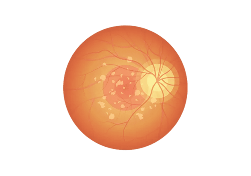
Macular degeneration is a disease that causes vision loss due to degeneration of the macula, located at the center of the retina.
The macula is the most critical part of the eye that enables us to see objects clearly, distinguish colors, and read text.
When abnormalities occur here, the central vision becomes blurred or distorted, causing significant inconvenience in daily life.
Macular degeneration is the leading cause of blindness worldwide, and in Korea, the number of patients is rapidly increasing alongside the aging population.
While it primarily affects those over 50, there is a recent trend of increasing cases among patients in their 40s.
Types of Macular Degeneration
Dry (non-exudative) macular degeneration
It accounts for approximately 85 to 90 percent of all macular degeneration cases.
It progresses gradually as waste products called drusen accumulate in the macula.
In the early stages, symptoms are often absent or mild, but if left untreated, it can progress to the wet form.
Exudative Macular Degeneration
Abnormal new blood vessels grow beneath the macula, causing bleeding and swelling.
Although it affects only 10–15% of cases, it carries a high risk of blindness,
and its rapid progression makes early treatment essential.
Symptoms of Macular Degeneration
Macular degeneration is a slowly progressing disease in which the macula,
the area responsible for central vision, gradually deteriorates.
In the early stages, patients often do not experience
noticeable discomfort.
Straight lines appear curved.
This is the most characteristic symptom.
Straight lines appear distorted and wavy, such as door frames, tile grout lines, and lines of text in books.
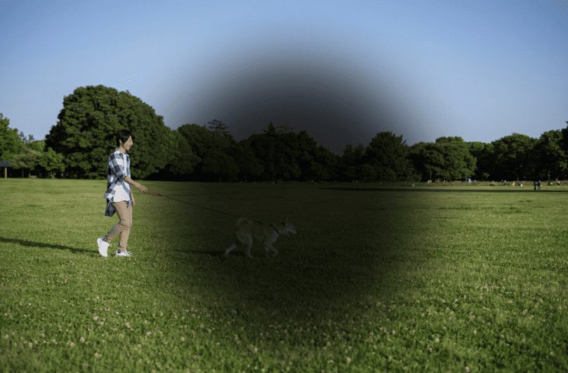
Blurred or darkened central vision
The area you want to see, such as the center of a person's face or text, appears blurry or obscured by black spots.
Difficulty distinguishing colors
Colors appear faded, and contrast sensitivity decreases, making it difficult to distinguish similar colors.
Sudden vision loss in dark places
In dimly lit environments, it becomes difficult to recognize objects,
and even when moving to a brighter area, it takes a long time to adjust.
Double vision
When looking with one eye versus the other, objects may appear to be different sizes.
Sudden vision loss
In particular, with wet macular degeneration, bleeding can cause a sudden decline in vision within days.
Emergency treatment is required in such cases.
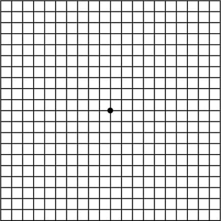
Macular Degeneration Self-Diagnosis
Using the Amsler grid test below,you can easily determine whether macular degeneration is progressing.
1. Wear the glasses or contact lenses you use in bright lighting during the examination. 2. Cover one eye and look at the round dot on the grid from a distance of about 30 cm. 3. Fix your gaze on the round dot in the center, then remember how the lines appear. 4. Examine the other eye using the same method.
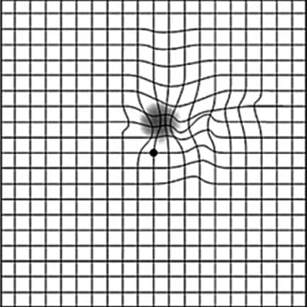
If you experience the following symptoms, you need to visit an ophthalmologist..
The line appears to curve in a wave-like pattern.
The round dot in the center is not clearly visible.
The middle part of the line appears broken.
A portion of the line appears blurred or distorted.
I see a dot that isn't in the picture.
Causes of Macular Degeneration
Aging
This is the primary cause. As we age, the function of retinal pigment epithelial cells declines and waste products accumulate, leading to progressive macular damage.
The incidence rate increases sharply after age 50.
Genetic factors
Having a family member with macular degeneration increases your risk of developing the condition by 3 to 4 times.
Regular checkups are especially important if you have a direct family member with a history of the disease.
Smoking
Smokers have a 2 to 5 times higher incidence of macular degeneration compared to non-smokers.
Smoking damages retinal blood vessels and increases oxidative stress,
promoting macular damage.
UV exposure
Prolonged exposure to strong sunlight causes cumulative damage to retinal cells, increasing the risk of macular degeneration.
Cardiovascular disease and hypertension
Poor vascular health can also affect blood flow to the retina,
increasing the risk of developing macular degeneration.
Obesity and high-fat diets
Research indicates that dietary habits characterized by obesity and
high saturated fat intake
are associated with the progression of macular degeneration.
Diagnosis/Examination of Macular Degeneration
Fundus examination
After dilating the pupils, the retina and macula are directly observed using a special lens.
This allows for the detection of drusen, hemorrhages, and pigment abnormalities.
Fundus photography
We take retinal photographs to document changes in the macula
and monitor them over time.
Optical Coherence Tomography (OCT)
High-resolution imaging of retinal cross-sections
enables precise assessment of macular edema, retinal layer damage, and the presence of neovascularization.
Prevention Guidelines for Macular Degeneration
No Smoking
Smoking is the strongest risk factor for macular degeneration. Quitting smoking alone can significantly reduce the risk.
UV protection
Wear sunglasses and a hat when going outside to protect your eyes.
Diet Management
Consume foods rich in lutein and omega-3 fatty acids, such as dark green leafy vegetables (spinach, kale, broccoli), oily fish (mackerel, salmon), and nuts.
Cardiovascular Health Management
Managing hypertension, hyperlipidemia, and obesity can also reduce the risk of macular degeneration.
Regular checkup
If you are 50 or older, it is important to undergo an annual fundus examination for early detection.
If you have a family history, screening is recommended starting in your 40s.
FAQ
Q. Does macular degeneration cause blindness?
Macular degeneration can damage central vision, but peripheral vision is usually preserved, so it rarely leads to complete blindness.
However, without early detection and appropriate treatment, it can lead to severe vision loss, making prompt action crucial.
Q. Can macular degeneration occur in both eyes?
Yes, if you have macular degeneration in one eye, there is a 40 to 50 percent chance it will develop in the other eye within five years.
Therefore, it is important to have regular eye exams for both eyes.
Q. Is macular degeneration hereditary?
There is a genetic component. Having a family member with macular degeneration increases your risk of developing it by 3 to 4 times.
If you have a direct family member with the condition, it is recommended to begin regular screenings starting in your 40s.
Q. Can macular degeneration occur at a young age?
Age-related macular degeneration primarily occurs in individuals over 50, but macular disease can also develop at a younger age due to genetic factors or high myopia.
If you experience visual field abnormalities, it is advisable to get examined regardless of age.
Learn more
Latest Information on Retinal Diseases
Check out the blog below for more information on retinal diseases.
 English (UK)