Retinal Vascular Occlusion

What is Retinal Vascular Occlusion?

Retinal vascular occlusion is a condition characterized by the obstruction of blood vessels that supply or drain the retina, resulting in impaired vision.

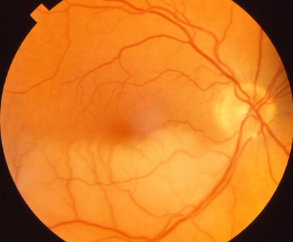
The retina is a critical tissue that converts incoming light into electrical signals for transmission to the brain, making a continuous blood supply essential.

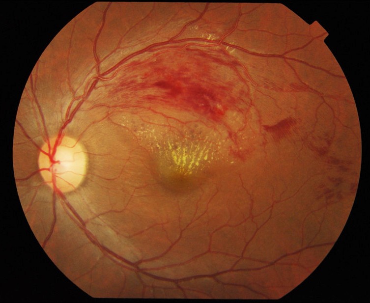
When an occlusion occurs, the retinal tissue is deprived of oxygen and nutrients, leading to damage, hemorrhage, or edema that adversely affects visual acuity.

Types of Retinal Vascular Occlusion

Retinal Artery Occlusion

Retinal Artery Occlusion is a condition involving the blockage of the arteries that supply blood to the retina. When an artery is obstructed, the retina is deprived of essential oxygen and nutrients, which can lead to a sudden and severe decline in visual acuity. 

A hallmark of retinal artery occlusion is the rapid onset of vision loss, typically occurring in only one eye.

Retinal Vein Occlusion

Retinal Vein Occlusion is a condition characterized by a blockage in the veins that drain blood away from the retina.

This obstruction interferes with intraretinal blood circulation, potentially leading to edema (swelling), hemorrhage (bleeding), and vision loss.

RVO is more prevalent than retinal artery occlusion and is highly associated with systemic conditions such as hypertension and diabetes.

Symptoms of retinal vascular occlusion

Retinal vascular occlusion symptoms typically manifest rapidly and can have a significant impact on visual acuity.

Sudden Decline in Visual Acuity

Patients may experience sudden blurring or

a rapid loss of vision, typically occurring in one eye (unilateral).

Partial vision obstruction

Part of my vision feels blurry or obscured.

A section of my vision often darkens

or disappears.

Presence of Visual Floaters or Photopsia

Patients may experience the sensation of small spots

or flashes of light drifting across their field of vision.

Transient Monocular Vision Loss

Vision is lost for a duration ranging from several seconds to a few minutes,

followed by spontaneous recovery.

Visual Distortion

If the macula is affected, straight lines may appear wavy, or objects may seem distorted or misshapen.

Blurred or Hazy Vision

When accompanied by macular edema, the patient's

overall vision may appear blurred or hazy, as if looking through a thick fog.

Causes of Retinal Vascular Occlusion

Hypertension

High blood pressure can damage the blood vessels in the retina

and cause them to narrow or become blocked.

Diabetes

Diabetes can damage blood vessels

and cause retinal vascular occlusion.

Atherosclerosis

Atherosclerosis is a condition where blood vessels thicken and stiffen,

leading to narrowing or blockage of the vessels, which can cause retinal vascular occlusion.

Hyperlipidemia

High cholesterol and triglycerides promote atherosclerosis and increase the risk of blood clot formation.

Heart Disease and Arrhythmia

Blood clots formed in conditions such as atrial fibrillation

can travel to the retinal artery

and cause its occlusion.

Other factors

Obesity, obstructive sleep apnea, and dehydration

are also known risk factors.

Retinal Vascular Occlusion Diagnosis/Examination

Safety Inspection

Optical Coherence Tomography (OCT)

Detailed examinations such as retinal angiography

Treatment for Retinal Vascular Occlusion

Intravitreal Antibody Injection for Reducing Macular Edema and Preventing Neovascularization

Local retinal laser treatment to prevent neovascularization

Prevention Guidelines for Retinal Vascular Occlusion

Blood Pressure Management

High blood pressure is the most important risk factor. Regularly measure your blood pressure and keep it within the target range.

Diabetes Management

Diabetes patients must strictly manage their blood sugar levels and undergo regular eye exams to monitor the condition of their retinas.

Cholesterol Management

Preventing and treating hyperlipidemia is important.

Blood lipid levels should be managed through regular exercise and dietary therapy.

Regular Checkup

High-risk individuals undergo regular fundus examinations to monitor the condition of their retinal blood vessels.

If retinal vascular occlusion occurs in one eye, the other eye must also be closely monitored.

FAQ

Retinal artery occlusion, particularly central retinal artery occlusion, is an ophthalmic emergency. Treatment must be administered within 90 minutes to several hours after onset to minimize retinal damage.

Retinal vein occlusion is not as urgent as artery occlusion, but prompt diagnosis and treatment significantly influence visual prognosis. Therefore, prompt ophthalmic evaluation is essential upon symptom onset.

Yes, retinal artery occlusion in particular often occurs through mechanisms similar to stroke (thrombosis, embolism).

Patients with retinal artery occlusion have a high risk of future stroke and myocardial infarction, making cardiovascular screening and preventive management essential.

Retinal vein occlusion is also strongly associated with cardiovascular risk factors.

It primarily occurs in individuals over 50, but it can also affect younger people.

In younger patients, specific underlying causes such as blood clotting disorders, inflammatory vascular diseases, or heart valve diseases may be present, necessitating additional testing.

The underlying causes of retinal vascular occlusion—hypertension, diabetes, and atherosclerosis—affect both eyes and the blood vessels throughout the body.

It can also occur in the other eye, and the risk of cardiovascular disease (stroke, myocardial infarction) increases, so managing overall health and undergoing regular checkups is important.

Learn more

Latest Information on Retinal Diseases

Check out the blog below for more information on retinal diseases.

KakaoTalk

Channel

YOUTUBE

Channel